Innovative Technique to Distinguish Healthy from Cancerous Cells
Synopsis
A team from Tokyo Metropolitan University has discovered a new method to differentiate between cancerous and healthy cells by tracking their movements, achieving 94% accuracy. This technique also opens doors for research into cellular functions related to motility, such as tissue healing.
Key Takeaways
New method identifies cancerous vs healthy cells.
94% accuracy in differentiation achieved.
Technique could aid in tissue healing research.
Uses phase-contrast microscopy, a label-free method.
Focus on cell motility and migration patterns.
Tokyo, April 19 (NationPress) A team of researchers from Tokyo Metropolitan University in Japan announced on Saturday that they have discovered a method to utilize the movement of unmarked cells to differentiate between cancerous and healthy cells.
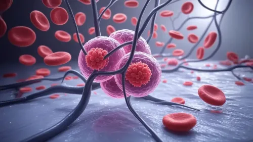
By examining malignant fibrosarcoma cells alongside healthy fibroblasts on a petri dish, they found that tracking and analyzing their trajectories can achieve differentiation with an impressive accuracy of up to 94 percent.
This method, beyond its diagnostic potential, may provide insights into cellular functions associated with motility, such as tissue repair, as detailed in their study published in the journal PLOS One.
Led by Professor Hiromi Miyoshi, the research team developed a technique for tracking cells using phase-contrast microscopy, a widely used method for cell observation.
This microscopy technique is completely label-free, allowing cells to move in a manner closer to their natural state, unaffected by the optical characteristics of the plastic petri dishes used for imaging.
Through advanced image analysis, they successfully captured the trajectories of numerous individual cells, concentrating on attributes like migration speed and the curvature of their paths, all of which reflect subtle distinctions in movement and deformation.
As a validation, they compared healthy fibroblast cells—essential components of animal tissues—with malignant fibrosarcoma cells, which are cancerous and originate from fibrous connective tissue.
The researchers demonstrated that the cells exhibited distinct migration patterns, characterized by the “sum of turn angles” (the curvature of their paths), the occurrence of gentle turns, and their speed. Remarkably, by analyzing both the sum of turn angles and the frequency of shallow turns, they were able to predict if a cell was cancerous with an accuracy of 94 percent.
This innovative work not only offers a groundbreaking method for identifying cancer cells but also holds potential applications for studying various biological functions related to cell motility, such as wound healing and tissue regeneration, the study reported.