Recent Research on Brain's Visual Areas Offers Insights for AI Development
Synopsis
A Columbia University study indicates that the brain's visual regions are crucial for interpreting information, which may lead to the development of more adaptable AI systems. The findings challenge previous beliefs about sensory areas and highlight the brain's flexibility in decision-making.
Key Takeaways
The brain's visual regions significantly influence information processing.
Flexibility in the brain allows for real-time adaptations in decision-making.
Research utilizes fMRI to observe brain activity during categorization tasks.
Visual cortex adjustments provide insights for AI system design.
Study challenges traditional views on sensory area functions.
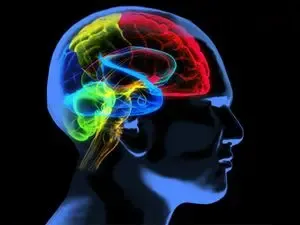
New Delhi, April 20 (NationPress) A study conducted by Columbia University’s School of Engineering in the United States has revealed that the brain’s visual regions actively contribute to processing information, which could aid in the creation of more adaptive AI systems.
Significantly, the brain's interpretation of information is influenced by other ongoing cognitive tasks.
Published in the journal Nature Communications, the research led by biomedical engineer and neuroscientist Nuttida Rungratsameetaweemana presents compelling evidence that early sensory systems are involved in decision-making and can adapt instantaneously.
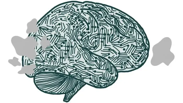
This research opens new avenues for developing AI systems capable of adjusting to unforeseen circumstances.
The findings contradict the traditional perspective that early sensory regions merely “observe” or “record” visual stimuli. Instead, the human brain’s visual system actively modifies its representation of the same object based on the current objectives.
Even within visual areas closely associated with raw data entering the eyes, the brain demonstrates the ability to adjust its interpretations and responses according to the task at hand.
“This provides a novel perspective on brain flexibility and inspires ideas for constructing more adaptive AI systems modeled after these neural frameworks,” stated Nuttida.
While most prior research focused on how individuals learn categories over time, this study emphasizes flexibility: How does the brain swiftly transition between diverse methods of organizing identical visual data?
The research team utilized functional magnetic resonance imaging (fMRI) to monitor brain activity as participants categorized shapes under changing rules.
This approach enabled the researchers to ascertain whether the visual cortex adjusted its representation of shapes based on the defined categories.
Data analysis employed computational machine learning techniques, including multivariate classifiers.
Activity within the visual system—encompassing the primary and secondary visual cortices, which process data directly from the eyes—varied with nearly every task.
The visual cortices restructured their activity based on the decision rules employed by participants, as evidenced by distinctive brain activation patterns when shapes approached the ambiguous boundary between categories.
These were the most challenging shapes to distinguish, indicating that additional processing is particularly beneficial in such scenarios.
“We observed clearer neural patterns in the fMRI data when participants performed better on the tasks, suggesting that the visual cortex may assist in solving flexible categorization challenges,” noted Nuttida.
The team is beginning to investigate how these concepts could be applied to artificial systems.