Toxic Metal Nanoparticles from MRI Scans Can Penetrate Human Tissue: Research
Synopsis
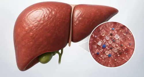
A recent study reveals that gadolinium, a toxic rare earth metal used in MRI scans, can form nanoparticles in human tissues through the action of oxalic acid found in various foods, raising concerns about health risks.
Key Takeaways
Gadolinium can create nanoparticles in the body.
Oxalic acid from foods aids this process.
Potential health risks include nephrogenic systemic fibrosis .
Gadolinium particles can linger in the body for years.
Recommendations regarding vitamin C and MRI scans are anticipated.
New Delhi, April 6 (NationPress) Researchers have discovered that a harmful rare earth metal utilized in MRI scans can produce nanoparticles of this metal within human tissues.
Investigators from the University of New Mexico in the United States, examining the health hazards linked to gadolinium, a toxic rare earth metal employed in MRI procedures, have uncovered that oxalic acid, a compound present in various foods, can lead to the formation of metal nanoparticles in human tissues.
In a recent article published in the journal Magnetic Resonance Imaging, the research team, headed by Brent Wagner, a professor in the Department of Internal Medicine, aimed to clarify the development of these nanoparticles, which have been connected to serious health issues in the kidneys and other organs.
“The most severe condition induced by MRI contrast agents is nephrogenic systemic fibrosis,” he stated. “There have been fatalities after just a single administration.”
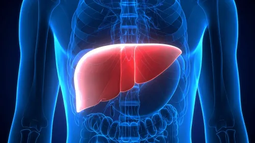
This condition could lead to a thickening and hardening of the skin, heart, and lungs, and result in painful joint contractures.
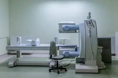
Gadolinium-based contrast agents are administered prior to MRI scans to enhance image clarity, as noted by Wagner.
This metal is typically securely attached to other molecules and is expelled from the body, with most individuals experiencing no negative effects.
However, past studies have indicated that even in asymptomatic individuals, gadolinium particles can be detected in the kidneys and brain, as well as in blood and urine years post-exposure.
Researchers are confronted with complex questions: Why do some individuals fall ill while most remain unaffected, and how do gadolinium particles detach from their accompanying molecules in the contrast agent?
“Almost half of the patients had only a single exposure, indicating that there is something that enhances the disease signal,” Wagner mentioned.
Wagner’s team concentrated on oxalic acid, found in numerous plant-based foods such as spinach, rhubarb, most nuts and berries, and chocolate, because it binds with metal ions.
This process can result in the formation of kidney stones, which occur when oxalate binds with calcium. Additionally, oxalic acid is also generated in the body when individuals consume foods or supplements containing vitamin C.
In laboratory tests, the researchers observed that oxalic acid prompted tiny quantities of gadolinium to precipitate from the contrast agent and form nanoparticles that subsequently infiltrated the cells of various organs.
This discovery suggests a potential method to reduce some of the risks linked with MRI scans.
“I would avoid taking vitamin C if I needed an MRI with contrast due to the metal's reactivity,” Wagner advised. “I am hopeful that we are progressing toward recommendations to assist these patients.”