NIT Rourkela Develops Natural Bioink for Bone Tissue 3D Bioprinting
Synopsis
Researchers at NIT Rourkela have innovatively developed a natural bioink aimed at improving 3D bioprinting of bone-like structures, tackling challenges in bone grafting and supporting regeneration.
Key Takeaways
Bioink made from natural materials.
Addresses bone grafting challenges.
Supports bone regeneration.
Stays liquid at room temperature.
Enhances treatment efficiency.
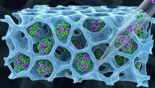
New Delhi, March 28 (NationPress) Scientists at the National Institute of Technology (NIT) Rourkela have created a bioink derived from natural substances to enhance 3D bioprinting of bone-like structures.
This innovative biocompatible bioink is tailored to tackle issues related to bone grafting and implants, which are frequently employed to remedy bone defects resulting from injuries or illnesses. Additionally, it fosters bone regeneration.
“This study adds to the expanding area of 3D bioprinting by providing a bioink that is completely natural, straightforward to use, and capable of facilitating bone regeneration,” stated Prof. Devendra Verma, Associate Professor in the Department of Biotechnology and Medical Engineering at NIT Rourkela.
3D bioprinting is increasingly considered as a viable alternative for bone repair, utilizing bio-inks that incorporate cells and supportive biomaterials to print bone-like structures.
Nevertheless, a significant hurdle with current bioinks is that the printed tissues require a controlled environment for cell development to form functional bone before being applicable for treatment. This slows down the process and complicates clinical implementation.
Conversely, the newly formulated bioink remains liquid at room temperature but swiftly transitions to a gel when influenced by body temperature and pH levels.
“This feature enables it to be printed directly at the site of injury, meaning the material can be applied on the injury instead of being separately printed and subsequently implanted. This method streamlines the procedure and enhances treatment efficiency,” the researchers noted in their publication in the Journal of Biomaterials Science and Carbohydrate Polymers.
The bioink comprises chitosan, gelatin, and nanohydroxyapatite, all of which are biocompatible and widely used in biomedical applications. These components closely mimic the natural constituents of bone, establishing an optimal environment for bone regeneration.
Additionally, the bioink encourages stem cell growth and their transformation into bone cells, further aiding in new bone formation.
The integration of specialized nanofibers boosts cell attachment and proliferation, which is crucial for the healing process.
“Further investigations and clinical trials will clarify its effectiveness in practical applications, paving the way for its integration into orthopedic and reconstructive surgical procedures,” Verma mentioned.
The research team is now set to test the bioinks on appropriate animal models and develop a scalable production method to support clinical trials.